January 16, 2026
5 min read
A 43-year-old woman with a past medical history significant for HIV complicated by cryptococcal meningitis and seizures presented to the ED after suffering a seizure and syncope.
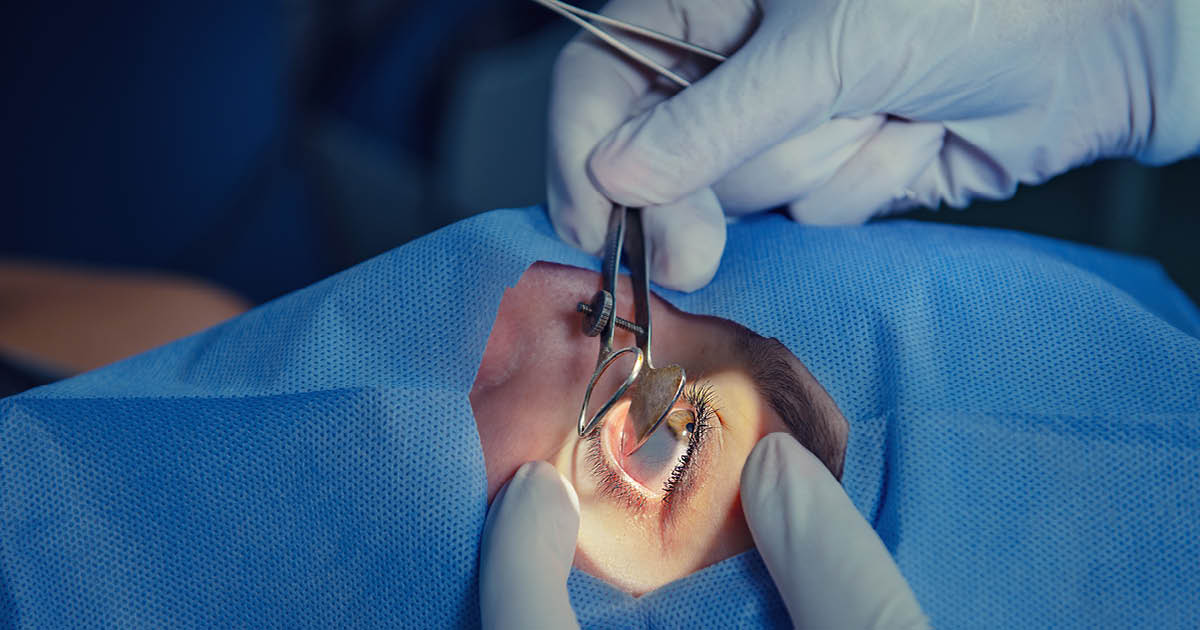
The following day she started complaining of left shoulder pain, and an orthopedic evaluation revealed posterior shoulder dislocation. As the patient was unable to tolerate an axillary view, anterior-posterior and scapular-Y radiographs were performed (Figures 1 and 2), demonstrating posterior dislocation of the shoulder. This was later confirmed with a CT scan of the left shoulder, which also demonstrated a reverse Hill-Sachs and reverse bony Bankart defect (Figures 3 and 4).

Source: Kevin Credille, MD; Jennifer Liu, MD; Patrick C. McCulloch, MD; and Joshua Woody, MD
What are the best next steps in management of this patient?
See answer below.

Source: Kevin Credille, MD; Jennifer Liu, MD; Patrick C. McCulloch, MD; and Joshua Woody, MD

Source: Kevin Credille, MD; Jennifer Liu, MD; Patrick C. McCulloch, MD; and Joshua Woody, MD
While admitted on the floor, the patient declined reduction attempts without sedation and was successfully closed reduced in the OR. She was seen by neurology to ensure optimal management of her epilepsy. After discharge, she was subsequently lost to follow-up and returned 4 months later with a locked posterior dislocation (Figure 5).

Source: Kevin Credille, MD; Jennifer Liu, MD; Patrick C. McCulloch, MD; and Joshua Woody, MD

Source: Kevin Credille, MD; Jennifer Liu, MD; Patrick C. McCulloch, MD; and Joshua Woody, MD
She reported recurrent dislocation approximately 3 weeks after the initial reduction, without trauma or seizure, accompanied by chronic shoulder pain. After that visit, outpatient CT demonstrated remodeling and resorption of the anterior humerus and posterior glenoid (Figure 6), while MRI revealed posterior labral tearing and a full-thickness subscapularis tear (Figures 7 and 8). After careful review of the advanced imaging at the next clinic visit with the patient, they elected to undergo surgical correction.

Source: Kevin Credille, MD; Jennifer Liu, MD; Patrick C. McCulloch, MD; and Joshua Woody, MD
Surgical technique
The patient received standard preoperative antibiotics, an interscalene block and general anesthesia, and was positioned in the beach chair. A standard deltopectoral approach was used. During exposure, full-thickness tearing of the superior border of the subscapularis was encountered. Using this interval, the upper two-thirds was taken down and retaining sutures were placed. Subacromial decompression, coracoacromial ligament release and subacromial bursectomy were then performed. After appropriate exposure, the humeral head was pushed posterior with lateral distraction with a bone hook to unlock and reduce the shoulder. The long head of biceps was tenodesed to the pectoralis.

Source: Kevin Credille, MD; Jennifer Liu, MD; Patrick C. McCulloch, MD; and Joshua Woody, MD
Attention was turned to the posterior reverse bony Bankart. A distal tibia allograft was sized, pre-drilled and pulse irrigated. Through a posterior cannula, the glenoid defect was prepared with a rasp and elevator. The graft was secured to the posterior glenoid with two fully threaded screws. The arm was taken through range of motion and was noted to be stable. Intraoperative fluoroscopy was used to confirm position.

Source: Kevin Credille, MD; Jennifer Liu, MD; Patrick C. McCulloch, MD; and Joshua Woody, MD
Humeral reconstruction was performed using an osteochondral allograft. The appropriate sizing guide was selected to contain the defect. A guide pin was placed, and the recipient socket was reamed. The osteochondral allograft was sized, prepped and pulse irrigated to remove marrow elements on the back table. It was then press fit into the recipient socket with excellent purchase (Figure 9). Two standard headless compression screws were used to gain additional purchase given her seizure disorder. The shoulder was again taken through range of motion and found to be stable.

Source: Kevin Credille, MD; Jennifer Liu, MD; Patrick C. McCulloch, MD; and Joshua Woody, MD
The subscapularis was repaired to the lesser tuberosity footprint using two medial all-suture anchors and a lateral row of anchors. The incision was closed in layers in standard fashion.
Postoperative course
The patient has followed up at 1 month and 3 months without recurrent dislocation or complications. She has consistently engaged with physical therapy and adhered to her rehabilitation protocol.

Source: Kevin Credille, MD; Jennifer Liu, MD; Patrick C. McCulloch, MD; and Joshua Woody, MD
Three-month radiographs demonstrated maintained reduction, well-positioned grafts and no hardware failure (Figures 10 and 11). At the time of that visit, which is her most recent range of motion check, she achieved 82° abduction, 97° forward flexion and 35° external rotation. During 6 months of consistent therapy, progressive improvements in range of motion and strength have been noted at each visit.

Source: Kevin Credille, MD; Jennifer Liu, MD; Patrick C. McCulloch, MD; and Joshua Woody, MD
Discussion
Posterior shoulder instability is more common in seizure disorder patients due to the relative strength of the internal rotators of the shoulder (subscapularis, pectoralis, latissimus and teres major) compared with the external rotators. Sudden, forceful muscle contractions occur during seizures that can lever the humeral head posteriorly out of the glenoid, frequently resulting in reverse Hill-Sachs lesions, posterior labral tears and reverse bony Bankart lesions. Posterior dislocations are estimated to account for nearly 36% of seizure related dislocations, while in the general population they only comprise 2% to 4% of dislocations. Delayed presentation, recurrent dislocations, soft tissue compromise and progressive bone loss can result in joint locking, as observed in our patient 4 months after initial reduction.

Source: Kevin Credille, MD; Jennifer Liu, MD; Patrick C. McCulloch, MD; and Joshua Woody, MD
Even modest posterior glenoid bone loss, as little as 11% of the glenoid diameter, increases the risk for recurrence after isolated soft tissue repair without bony augmentation, highlighting the importance of addressing concomitant bony and soft tissue pathology at the time of surgery. Surgical techniques for addressing posterior glenoid bone loss include distal tibial allograft, iliac crest autograft and posterior glenoid osteotomy. Distal tibial allograft offers several advantages:
- provides a large, structurally robust graft for posterior augmentation;
- avoids donor site morbidity associated with autografts;
- closely matches the native concave glenoid articular surface; and
- has demonstrated low rates of recurrent instability and graft resorption.
In cases of combined humeral and glenoid defects, dual allograft reconstruction using humeral osteochondral allograft and posterior glenoid osteochondral allograft allows anatomic reconstruction of both articulating surfaces. Although osteochondral allograft for reverse Hill-Sachs lesions is not commonly performed, it effectively restores the humeral articular surface, preserves motion and features good outcomes in patients with locked posterior dislocations. In patients who may require dual allograft reconstruction and the addition of a humeral osteochondral allograft, as seen in this case, successful postoperative outcomes are achievable with appropriate patient selection, meticulous surgical technique and adherence to postoperative rehabilitation protocols.
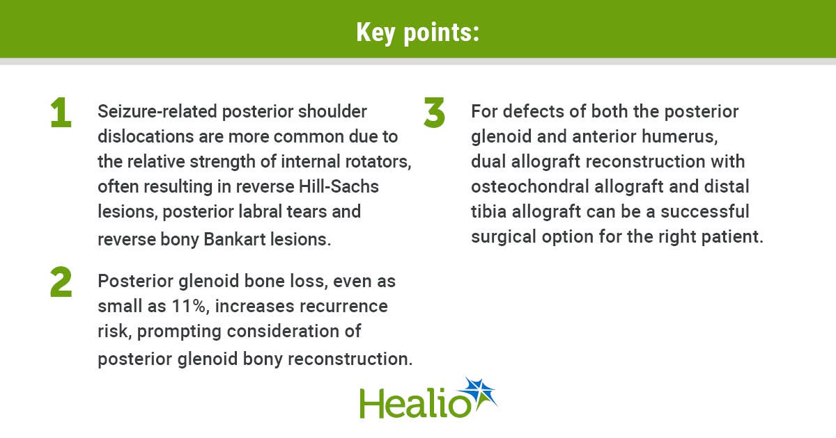
Key points:
- Seizure-related posterior shoulder dislocations are more common due to the relative strength of internal rotators, often resulting in reverse Hill-Sachs lesions, posterior labral tears and reverse bony Bankart lesions.
- Posterior glenoid bone loss, even as small as 11%, increases recurrence risk, prompting consideration of posterior glenoid bony reconstruction.
- For defects of both the posterior glenoid and anterior humerus, dual allograft reconstruction with osteochondral allograft and distal tibial allograft can be a successful surgical option for the right patient.
For more information:
Kevin Credille, MD; Jennifer Liu, MD; Patrick McCulloch, MD; and Joshua T. Woody, MD, can be reached at Houston Methodist Hospital in Houston, Texas. Credille’s email: kcredille@houstonmethodist.org. Liu’s email: jwliu@houstonmethodist.org. McCulloch’s email: pcmcculloch@houstmethodist.org. Woody’s email: jtwoody@houstonmethodist.org.
Edited by Mitchell F. Bowers, MD, and Jennifer Liu, MD. Bowers is a chief resident in orthopedic surgery at Vanderbilt University Medical Center. He will be pursuing a spine surgery fellowship at the Leatherman Spine Institute following residency completion. Liu is a chief resident in orthopedic surgery at Houston Methodist Hospital. She will be pursuing an adult reconstruction fellowship at the University of California San Francisco following residency completion. For more information on submitting Orthopedics Today Grand Rounds cases, please email orthopedics@healio.com.
Sources/Disclosures
Source:
Expert Submission
References:
Disclosures:
McCulloch reports being a paid consultant for and receiving research support from Arthrex, and being on the editorial or governing board for Orthobullets.com. Credille, Liu and Woody report no relevant financial disclosures.
Ask a clinical question and tap into Healio AI’s knowledge base.
- PubMed, enrolling/recruiting trials, guidelines
- Clinical Guidance, Healio CME, FDA news
- Healio’s exclusive daily news coverage of clinical data
<


Leave a Reply