April 30, 2026
2 min read
Key takeaways:
- Machine learning was used to detect interstitial lung disease in patients with connective tissue disease.
- The data may enhance interpretation by physicians or radiologists. .
Researchers successfully used machine learning on lung ultrasound images to detect interstitial lung disease in patients with systemic sclerosis and idiopathic inflammatory myopathy, according to data published in Arthritis Care & Research.
“Beyond its accuracy, [lung ultrasound (LUS)] offers advantages such as low cost, accessibility, no radiation, and sustainability,” Robert M. Fairchild, MD, PhD, clinical chief of the division of immunology and rheumatology at Stanford University, and colleagues wrote. “However, operator dependence and variability in acquiring and interpreting LUS remain a potential drawback. This may explain limited adoption of LUS for [connective tissue disease (CTD)]-ILD screening despite evidence supporting its use.

Data derived from Fairchild RM, et al. Arthritis Care Res. 2026;doi:10.1002/acr.80054.
“In this context, applying artificial intelligence (AI) to LUS in ILD offers an opportunity to address two key issues,” they added. “First, AI can help independently validate the diagnostic sufficiency of pleural-based sonographic features in ILD. … Second, AI has the potential to reduce operator-dependent variability, a major barrier to clinical adoption, thereby supporting broader implementation of LUS as a scalable, accessible, and reliable modality for ILD detection in connective tissue diseases.”
In the current study, Fairchild and colleagues aimed to determine whether the application of deep learning, convolutional neural networks (CNN) to lung ultrasound could accurately detect ILD and its severity among patients with SSc or idiopathic inflammatory myopathy. The analysis included data for 3,920 ultrasound images from 140 individuals with SSc or idiopathic inflammatory myopathy plus ILD. Eligible participants had undergone paired lung ultrasound and chest CT.
The researchers used transfer learning to “fine tune” three pre-trained CNN architectures — InceptionV3, ResNet-50 and VGG-16 — and develop a de novo lightweight architecture called LUS-Net, they wrote. In all, 74 patients were assigned to a development test set while 66 were assigned to an independent test set.
According to the researchers, VGG-16 performed best in assessing patient-level data, with a sensitivity of 97.4% and a specificity of 92.6% (AUC = 0.972). VGG-16 also correlated strongly with pulmonary function test data and CT severity, according to the findings.
“Grad-CAM highlighted pleural features as the primary regions influencing model predictions,” Fairchild told Healio in an interview. “CNN performance matched or exceeded LUS-ILD-24 interpretation — a high-performing and validated human lung ultrasound interpretation criteria for ILD detection in systemic sclerosis and inflammatory myopathy.”
The researchers concluded that deep learning may successfully be applied to lung ultrasound for the detection of ILD. These data may enhance interpretation by physicians or radiologists, they added.
“Deep learning-based interpretation of lung ultrasound provides a promising and scalable solution for ILD detection in connective tissue disease,” Fairchild and colleagues wrote. “By combining the portability and safety of LUS with the power of CNNs, this approach has the potential to standardize interpretation, enhance understanding and transparency with explainable AI, and expand access to high-quality ILD screening and monitoring across a broad range of clinical settings.”
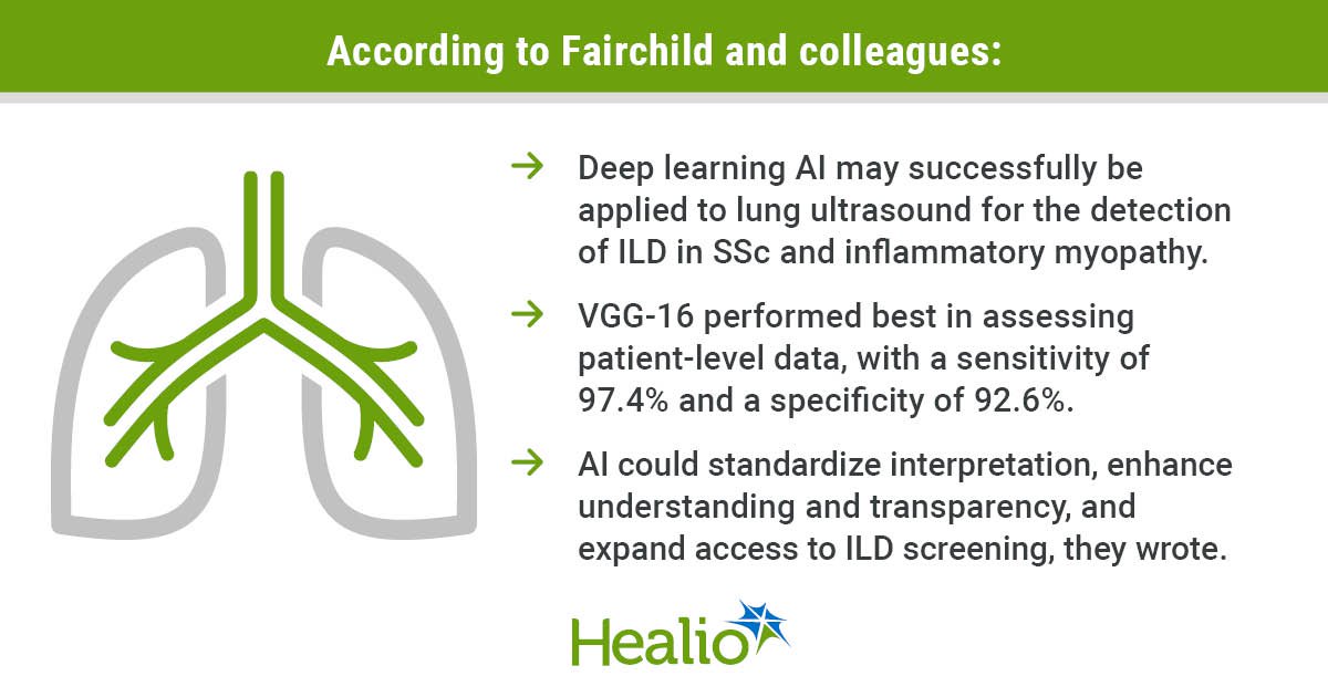
Deep learning AI may successfully be applied to lung ultrasound for the detection of ILD in SSc and inflammatory myopathy.
VGG-16 performed best in assessing patient-level data, with a sensitivity of 97.4% and a specificity of 92.6%.
AI could standardize interpretation, enhance understanding and transparency, and expand access to ILD screening, they wrote.
Ask a clinical question and tap into Healio AI’s knowledge base.
- PubMed, enrolling/recruiting trials, guidelines
- Clinical Guidance, Healio CME, FDA news
- Healio’s exclusive daily news coverage of clinical data
<


Leave a Reply