Key takeaways:
- Earlier diagnosis can improve outcomes in giant cell arteritis.
- Integrating imaging can improve diagnostic accuracy in vasculitis.
DESTIN, Fla. — Imaging analyses can improve diagnostic accuracy and confidence in patients with giant cell arteritis, according to data presented at the Congress of Clinical Rheumatology East.
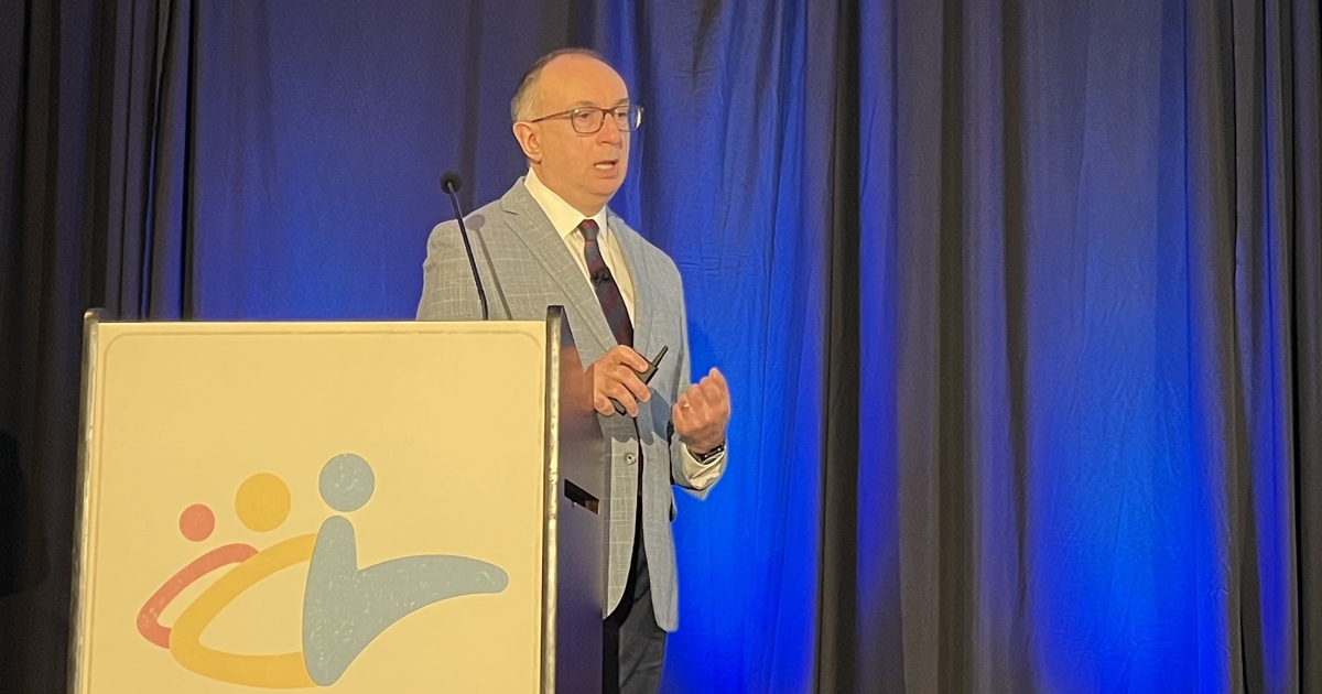
“The reason I am speaking on advances in GCA is that diagnosis and management continue to evolve, particularly with increasing use of advanced imaging,” Kenneth J. Warrington, MD, director of the vasculitis clinic, consultant in the division of rheumatology, and John F. Finn MN Arthritis Foundation Professor of Medicine at the Mayo Clinic College of Medicine and Science, told Healio. “Over the past few years, we have learned that integrating imaging earlier can improve diagnostic confidence.”

“The reason I am speaking on advances in GCA is that diagnosis and management continue to evolve, particularly with increasing use of advanced imaging,” Kenneth Warrington, MD, told Healio. Image: Rob Volansky | Healio
Temporal artery ultrasound, temporal artery biopsy, MRI of the brain with vessel wall imaging, computed tomography angiography (CTA) of the chest or abdomen, magnetic resonance angiography (MRA) of the chest or abdomen, or fluorodeoxyglucose-positron emission tomography (FDG-PET)/CT scan all may be used in various patients and at various time points in the disease course, according to Warrington.
“Now we have a whole menu of tests we can use to evaluate patients,” he said in his presentation. “The question is, how do we choose.”
For example, temporal artery ultrasound can be effective but comes with a qualification.
“Patients with atherosclerosis may receive an erroneous GCA diagnosis,” Warrington said.
Meanwhile, vessel wall MRI is being used with increasing frequency.
“It is a powerful tool to know whether arteries are inflamed or not,” Warrington said. “It has great specificity and sensitivity.”
However, the issue with vessel wall MRI is availability. Many centers do not yet have this technology, Warrington said.
When imaging the chest or abdomen, CTA can show thickening of the vasculature, while MRA can show edemas, according to Warrington.
“CT can tell us something is going wrong, but it does not give us any information about whether inflammation is active or not,” he said. “MRI is better at doing that.”
FDG-PET imaging may save time and effort in patients who “have bounced around” with a negative biopsy or ultrasound, if they have had a fever or inflammation of unknown origin, or a suspected malignancy, according to Warrington.
“It can be helpful even in patients on treatment,” he said.
Regarding the choice between biopsy and ultrasound, Warrington noted that the American College of Rheumatology recommends biopsy, while European guidelines favor ultrasound. Practitioners are encouraged to follow the guidelines in their region and assess individual patients for the preferential test, he said.
“The main take-home message for rheumatologists is the importance of early, accurate diagnosis, along with thoughtful use of imaging, and temporal artery biopsy in some cases, as a complement to clinical assessment,” Warrington told Healio. “When we have imaging and histopathology, we get a much better picture of what is going on with our patients.”
For more information:
Kenneth J. Warrington, MD, can be reached at Warrington.Kenneth@mayo.edu.
Sources/Disclosures
Source:
Warrington K. Advances in the diagnosis and treatment of giant cell arteritis. Presented at The Congress of Clinical Rheumatology East; April 30-May 3, 2026; Destin, Florida (hybrid meeting).
Disclosures:
Warrington reports receiving clinical trial support from Bristol Myers Squibb and consulting fees from Amgen, AstraZeneca and Sanofi.
Ask a clinical question and tap into Healio AI’s knowledge base.
- PubMed, enrolling/recruiting trials, guidelines
- Clinical Guidance, Healio CME, FDA news
- Healio’s exclusive daily news coverage of clinical data
<


Leave a Reply