
January 19, 2026
2 min read
Key takeaways:
- OCT, topography, biometry, aberrometry and meibography can be invaluable for surgical planning.
- Cataract surgeons should be vigilant for dry eye symptoms to ensure patient satisfaction.
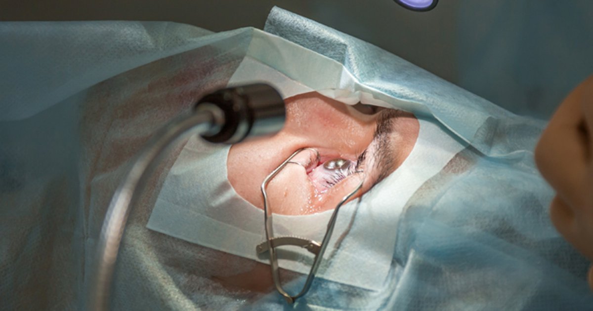
WAIKOLOA, Hawaii — Five diagnostic tools are critical to assist surgeons in choosing the most appropriate IOL for the patient and maintaining clinic flow and efficiency, according to a presenter.
At Hawaiian Eye 2026, Marjan Farid, MD, said macular OCT, topography/tomography, biometry, aberrometry and meibography are invaluable for surgical planning.

“There is a lot of information to think about when evaluating a patient for cataract surgery,” she said. “A lot of data goes in, and ultimately the processor is the surgeon’s mind in figuring out how to put all of this together to match the correct lens to our patient while maintaining clinic flow.”
Subtle macular and vitreous pathologies can easily be missed on a standard exam, Farid said, “and the vision sometimes doesn’t point to it, either. … Unless you do [macular OCT] before and show the patient they have a level of pathology in their retina, if it gets worse after surgery, which oftentimes it can, then the blame falls on the surgeon.”
Topography can be key to discovering hidden corneal irregularities, Farid said. Physicians should be sure to lift eyelids, view the superior cornea and plan an in-office excision if possible. Before IOL planning, topography should also be re-evaluated.
Today’s optical biometers have fourth-generation formulas that can provide axial length, K values and total corneal power, and aberrometers can help distinguish corneal and internal aberrations and serve as an educational tool for patients, she said.
“You can really show the patient what the difference is going to look like when you address the astigmatism,” Farid said.
Meibography and ocular surface disease assessment round out the diagnostics.
Farid emphasized that physicians need to remember that every patient is unique, and lifestyle questionnaires, a thorough medical history and biomicroscopy exams can also ensure the best outcomes for patients. She noted that it is advisable to conduct osmolarity and MMP-9 testing prior to exposing the eye to dilation, direct contact, vital dyes or bright lights.
Staff should be empowered to conduct testing so the surgeon can diagnose quickly and initiate ocular surface disease management where necessary, Farid explained.
“The number one, two and three causes of dissatisfied postoperative cataract patients are ocular surface disease and dry eyes,” she said. “Look for signs. Many patients will come in and are not complaining about the traditional symptoms of dry eye, but they have signs of ocular surface disease.”
Sources/Disclosures
Source:
Farid M. Top 5 diagnostics in cataract planning. Presented at: Hawaiian Eye 2026; Jan. 17-23, 2026; Waikoloa, Hawaii.
Disclosures:
Farid reports being a consultant or advisor to Alcon, AbbVie, Allergan, Aurion Biotech, Bausch + Lomb, Bio-Tissue, Carl Zeiss Meditec, CorneaGen, Glaukos, Harrow, Johnson & Johnson Vision Care, Kala Pharmaceuticals, Orasis Pharmaceuticals, Sight Sciences, Sun Ophthalmics, Tarsus Pharmaceuticals and Viatris.
Ask a clinical question and tap into Healio AI’s knowledge base.
- PubMed, enrolling/recruiting trials, guidelines
- Clinical Guidance, Healio CME, FDA news
- Healio’s exclusive daily news coverage of clinical data
<


Leave a Reply