Lindstrom’s Perspective
April 17, 2026
3 min read
Lindstrom’s Perspective
Click here to read the Cover Story, “Eye rubbing: Screen for it, address causes, make patients aware of consequences.”
In the past decade, we have learned much about keratoconus, and in this perspective, I will share a few learnings I find especially important.

I will disclose that, as a cornea specialist, the management of keratoconus was a significant part of my practice, and I participated in the Avedro, Glaukos and Epion clinical trials on collagen cross-linking and continue to consult widely in this field.
First, we have learned that keratoconus is much more prevalent than originally taught. Classic textbooks suggest that keratoconus is present in about one per 2,000 individuals. Current studies suggest that the prevalence of keratoconus is actually at least 5 to 10 times higher and in some Middle Eastern and African populations may occur in more than 1% of the population. Nearly all patients with keratoconus are myopic, and the increased interest in treating pediatric progressive myopia will help us diagnose keratoconus earlier. As more progressive myopes are captured in an eye care practice at age 4 to 5 years for treatment, it will be straightforward to screen for early keratoconus with retinoscopy, keratometry, pachymetry and corneal topography. This will allow us to diagnose keratoconus in the teens or early 20s when minimal regular and irregular astigmatism has developed and treat the disease immediately with corneal cross-linking. As discussed in the accompanying cover story, pediatric progressive myopes can be counseled to avoid eye rubbing, which may reduce keratoconus incidence. In addition, high-risk patients, including those with allergic eye disease or a positive family history, especially male patients, can be followed more closely. If we treat keratoconus before significant visual degradation occurs, nearly all newly diagnosed patents with keratoconus will be manageable with spectacles or soft contact lenses, and keratoplasty will rarely be required. In addition, the significant psychological issues and reduced quality of life associated with advanced keratoconus will be eliminated.
Second, we have learned that the diagnosis of keratoconus is itself an indication for immediate treatment in children and younger patients. Like progressive myopia, younger patients progress faster and can progress significantly in even a few months. Following patients until significant progression is confirmed or they become spectacle or even contact lens intolerant is not appropriate. The recent approval of epithelium-on cross-linking makes treatment of even the youngest patients possible under topical anesthesia with occasional oral sedation, similar to an office-based pediatric examination under anesthesia.
Third, collagen cross-linking is a disruptive innovation in the treatment of progressive keratoconus and when applied early can prevent the development of vision-disabling disease. Progressive pediatric myopia and keratoconus will often both be present, and it is appropriate to treat both simultaneously using behavioral modification including outside activities, no eye rubbing, specialized optical correction and topical low-dose atropine eye drops together with immediate corneal cross-linking.
Fourth, while the older patient with significant but still progressive keratoconus deserves timely treatment with collagen cross-linking, which over time can stabilize the disease and result in corneal flattening with reduced myopia and astigmatism along with improved uncorrected and best corrected vision, they also deserve to have refractive surgical options offered. Like the cataract/IOL patient of today in whom refractive cataract surgery options are available, there are also procedures that can reduce myopia and astigmatism and further enhance uncorrected and best corrected visual function for the patient with more advanced keratoconus. I call this refractive cross-linking, and at Minnesota Eye Consultants, we have found significant interest in corneal tissue addition keratoplasty (CTAK) or corneal allogenic intrastromal ring segments (CAIRS) and/or custom PRK with some truly amazing results. While early in adoption, I look forward to the day when nearly all patients with more advanced keratoconus can access treatment that not only prevents progression of their disease but also enhances their visual function.
Fifth, when a patient with keratoconus requires keratoplasty, there is growing evidence that deep anterior lamellar keratoplasty offers a better long-term outcome than penetrating keratoplasty, as most patients with keratoconus have a normal healthy endothelium. Unfortunately, most U.S. cornea surgeons continue to favor PK as it is technically easier. It is time for cornea specialists to develop the skills needed for DALK, just like they did over the last decade replacing PK with Descemet’s stripping endothelial keratoplasty and Descemet’s membrane endothelial keratoplasty for endothelial disease.
Finally, we have another disruptive innovation coming for the treatment of keratoconus, and that is topical eye drops that can induce collagen cross-linking. Early clinical trials are promising, and I see the day when, much like glaucoma, we will have a topical eye drop alternative to arrest progression.
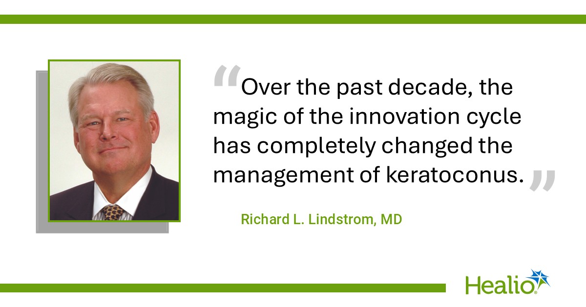
Over the past decade, the magic of the innovation cycle has completely changed the management of keratoconus. More patient and eye doctor education is needed to accelerate timely referral and early treatment with collagen cross-linking. In addition, patients deserve to know we can significantly enhance visual function when desired with CTAK, CAIRS and/or custom PRK.
For more information:
Richard L. Lindstrom, MD, can be reached at rllindstrom@mneye.com.
Lindstrom’s Perspective
Sources/Disclosures
Source:
Expert Submission
Disclosures:
Lindstrom reports having financial disclosures for Avedro, CorneaGen, Epion, Glaukos, iVeena, Minnesota Eye Consultants and Unifeye Vision Partners. Literature review augmented by Elicit, Google AI and Healio AI.
Ask a clinical question and tap into Healio AI’s knowledge base.
- PubMed, enrolling/recruiting trials, guidelines
- Clinical Guidance, Healio CME, FDA news
- Healio’s exclusive daily news coverage of clinical data
<


Leave a Reply