April 15, 2026
2 min watch
Key takeaways:
- EndoArt is an alternative to human endothelial tissue transplantation in complex cases.
- Studies show that it is safe, stable and effective.
HELSINKI — The EndoArt artificial endothelial layer showed positive results across multiple studies presented by specialists at the European Society of Cataract and Refractive Surgeons winter meeting.
“It looks like a contact lens, only you put it inside the eye and not on top of the cornea,” Ruth Lapid, MD, PhD, said.

Image: Michela Cimberle | Healio
Made of hydrophilic acrylic material, EndoArt (EyeYon Medical) adheres to the posterior surface of the cornea, acting as a barrier to the transfer of fluids. It is designed to treat chronic corneal edema in complex cases and serves as an alternative to human corneal transplantation.
Lapid discussed her 6-year experience with the implant at the University Medical Center Utrecht, Netherlands, and presented the results of a study of 27 patients with a follow-up from 1 to 6 years. All patients had complex presentations.
“Trauma, Fuchs’, glaucoma shunts, failed [penetrating keratoplasty] or [Descemet’s stripping endothelial keratoplasty], iatrogenic trauma — you name it, they have it,” she said.
However, results were rewarding, with corneal thickness decreasing by about 24% at 1 year and 28% at 3 years and mean visual acuity improving from counting fingers to 20/400.
“When I look at visual acuity, I think, ‘What a terrible outcome. Those people don’t see anything,’” Lapid said. “But the difference this little improvement makes in relation to moving in their houses, getting dressed alone and eating on their own is quite important for people who have bilateral pathology.”
In terms of adverse events, she reported two cases of failed adherence, two cases of epithelial fibrosis, two reactivations of herpetic keratitis and two cases of cystoid macular edema that resolved with treatment.
Sorcha Ní Dhubhghaill, MD, PhD, and Ruth Lapid, MD, PhD, discuss the EndoArt artificial endothelial layer, an alternative to human endothelial tissue transplantation.
“What we did not see, and that’s the better part, was pathological thinning of the cornea,” Lapid said. “So, once it’s in place, it seems to stick.”
Sorcha Ní Dhubhghaill, MD, PhD, on behalf of Ofer Daphna, MD, discussed results of more than 900 implantations performed to date at different sites, with a follow-up of more than 6.5 years. Central corneal thickness was reduced in 86% of eyes, vision improved in 60%, corneal clarity improved in 81%, and pain was reduced in 92%, with less dependency on bandage contact lenses observed.
Dhubhghaill specified that the most common indications for EndoArt are prior graft failure and previous glaucoma surgery, including filtering procedures and, more predominantly, drainage devices, both known to be poor prognostic indicators when using human tissue.
“These are the target groups where we believe that there is considerable added value with EndoArt,” Dhubhghaill said.
The researchers also compared normal-risk vs. high-risk cases for human tissue failure, with 70 cases and 76 cases included in each group, respectively.
“The question was, how does EndoArt perform in these two cases? And interestingly enough, there was no statistically significant difference between the groups in central corneal thickness or in corneal clarity outcomes,” Dhubhghaill said. “There were no serious adverse events in either group. The most common complication was re-bubbling … which was required in 55% of the higher-risk eyes and 45% of the normal eyes. Interestingly, explantation rates were higher in the normal-risk group, which was 12% vs. 4.5%.”
Natural tissue, whenever it can be used, gives the best outcomes, she noted, while EndoArt performs particularly well at the end of the biological line, when patients have limited options due to concurrent diseases or previous failed attempts with natural tissue, she said.
“Give the opportunity for real tissue first,” Dhubhghaill said.
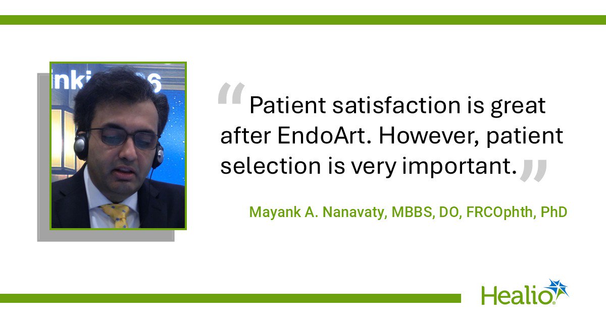
Another retrospective series of 14 patients implanted with EndoArt at a tertiary referral center in the U.K. was presented by Mayank A. Nanavaty, MBBS, DO, FRCOphth, PhD. Five of 14 eyes did not have previous keratoplasty, he said.
A mean 38% reduction in central corneal pachymetry was seen at 1 week, followed by a further reduction at 2 weeks, remaining stable afterward. At about 30 months, the cornea was still slightly thicker than normal, but this was a good result, Nanavaty said.
Considering that these patients have significant comorbidities, vision improved but remained within the low range because of concurrent conditions.
Hand movement vision was reduced from 50% of patients to 36%, and several patients transitioned from hand movement to measurable Snellen acuity of 6/24 to 6/30, “a meaningful gain for activities of daily living,” Nanavaty said.
The re-suturing rate was 35.7%, and no re-bubbling was performed. Nanavaty said that he and his group have stopped re-bubbling “because we know that re-bubbling is a time waster,” he said.
“We take five sutures at 2, 4, 6, 8 and 10 o’clock and keep them in for 3 months,” Nanavaty said. “We used to remove the sutures after 6 weeks, but that wasn’t enough. … We have also done safety net suturing in two patients.”
The transplant can significantly improve quality of life, and satisfaction is high with appropriate patient selection. More studies are needed to assess long-term durability, define patient selection criteria, better standardize the technique and evaluate quality of life through questionnaires, Nanavaty said.
“Patient satisfaction is great after EndoArt. However, patient selection is very important,” he said. “Visual acuity does improve, but it depends on the comorbidity of the patient.”
Sources/Disclosures
Source:
Healio Interviews
References:
- Dhubhghaill SN. EndoArt implantation in patients at high risk for human graft rejection. Presented at: European Society of Cataract and Refractive Surgeons winter meeting; March 6-8, 2026; Helsinki.
- Lapid R, et al. 6 year experience with the endothelial keratoprosthesis implantations in the Netherlands. Presented at: European Society of Cataract and Refractive Surgeons winter meeting; March 6-8, 2026; Helsinki.
- Nanavaty MA, et al. Clinical outcomes of artificial corneal endothelial transplant in a tertiary referral centre. Presented at: European Society of Cataract and Refractive Surgeons winter meeting; March 6-8, 2026; Helsinki.
Disclosures:
Dhubhghaill and Lapid report no relevant financial disclosures. Nanavaty reports receiving research grants from EyeYon Medical.
Ask a clinical question and tap into Healio AI’s knowledge base.
- PubMed, enrolling/recruiting trials, guidelines
- Clinical Guidance, Healio CME, FDA news
- Healio’s exclusive daily news coverage of clinical data
<


Leave a Reply